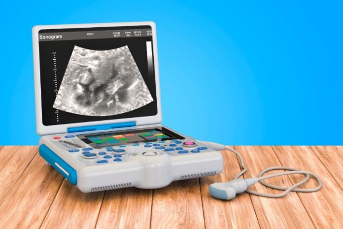
The field of veterinary medicine has seen significant advancements over the years, with technology playing a pivotal role in enhancing diagnostic capabilities and improving patient outcomes. Among the most transformative innovations is sound wave imaging, commonly known as ultrasound. This non-invasive diagnostic tool has revolutionized the way veterinarians diagnose and treat various conditions in animals. This article explores how sound wave imaging is changing veterinary diagnostics, highlighting its benefits, applications, and impact on veterinary practice.
Understanding Sound Wave Imaging
Sound wave imaging, or ultrasound, utilizes high-frequency sound waves to produce detailed images of internal structures within the body. Unlike X-rays, which use ionizing radiation, ultrasound is based on the principle of sound wave reflection. The sound waves are emitted by a transducer, penetrate the body tissues, and bounce back to the transducer when they encounter different tissues and organs. These reflected waves are then converted into real-time images displayed on a monitor.
Benefits of Sound Wave Imaging
1. Non-Invasive and Safe
One of the most significant advantages of ultrasound is its non-invasive nature. It does not require incisions or injections, making it a safer option for both animals and veterinarians. Additionally, because it does not use ionizing radiation, it poses no risk of radiation exposure to the patient, which is particularly important for repeated use in monitoring chronic conditions.
2. Real-Time Imaging
Ultrasound provides real-time images, allowing veterinarians to observe dynamic processes within the body, such as blood flow, organ movement, and fetal development. This capability is especially valuable in emergency situations where quick, accurate assessments are crucial.
3. Detailed Visualization
Sound wave imaging offers high-resolution images that provide detailed views of soft tissues, organs, and blood vessels. Techniques such as Doppler ultrasound enhance the visualization of blood flow, while contrast-enhanced ultrasound improves the detection of abnormalities like tumors.
4. Versatility
Ultrasound is a versatile diagnostic tool used to examine various body systems, including the cardiovascular, gastrointestinal, reproductive, and musculoskeletal systems. Its ability to provide comprehensive assessments makes it invaluable in diagnosing a wide range of conditions.
Applications in Veterinary Diagnostics
1. Cardiovascular Health
Doppler ultrasound is instrumental in assessing cardiovascular health in animals. It allows veterinarians to evaluate heart function, detect congenital heart defects, and monitor conditions like heart murmurs and arrhythmias. By visualizing blood flow patterns and measuring cardiac output, veterinarians can make informed decisions about treatment and management.
2. Abdominal Examinations
Ultrasound is widely used for abdominal examinations, providing detailed images of organs such as the liver, spleen, kidneys, and gastrointestinal tract. It aids in diagnosing conditions like liver disease, kidney stones, gastrointestinal obstructions, and tumors. Its non-invasive nature makes it ideal for monitoring chronic conditions and assessing treatment effectiveness.
3. Reproductive Health
In reproductive medicine, ultrasound is invaluable for monitoring pregnancy, detecting fetal abnormalities, and assessing reproductive organs. It allows veterinarians to determine the number of fetuses, estimate gestational age, and monitor the health of both the mother and the developing fetuses, ensuring successful pregnancies and timely interventions when necessary.
4. Musculoskeletal Evaluations
Ultrasound is also employed in evaluating musculoskeletal conditions. It helps diagnose ligament and tendon injuries, joint abnormalities, and soft tissue swellings. By providing real-time images of moving structures, ultrasound enables veterinarians to assess the extent of injuries and guide treatment plans effectively.
Impact on Veterinary Practice
The integration of sound wave imaging into veterinary practice has transformed diagnostic approaches and improved patient care. The ability to obtain detailed, real-time images non-invasively has led to earlier and more accurate diagnoses, enhancing patient outcomes. Additionally, the versatility of ultrasound allows for comprehensive assessments of various body systems, reducing the need for multiple diagnostic procedures and minimizing patient stress.
Furthermore, advancements in ultrasound technology have made it more accessible and affordable for veterinary clinics of all sizes. Portable ultrasound machines enable on-site imaging, facilitating timely care in emergency situations and during routine check-ups. This accessibility ensures that more animals can benefit from advanced diagnostic services, ultimately raising the standard of veterinary care.
Conclusion
Sound wave imaging has revolutionized veterinary diagnostics, offering numerous benefits for both patients and practitioners. Its non-invasive, real-time, and detailed imaging capabilities have transformed the way veterinarians diagnose and manage a wide range of conditions. As technology continues to advance, the future of veterinary medicine looks promising, with ultrasound playing a central role in improving the health and well-being of animal patients. By embracing these advancements, veterinary professionals can provide more effective and compassionate care, ensuring better outcomes for their furry, feathered, and scaled patients.


